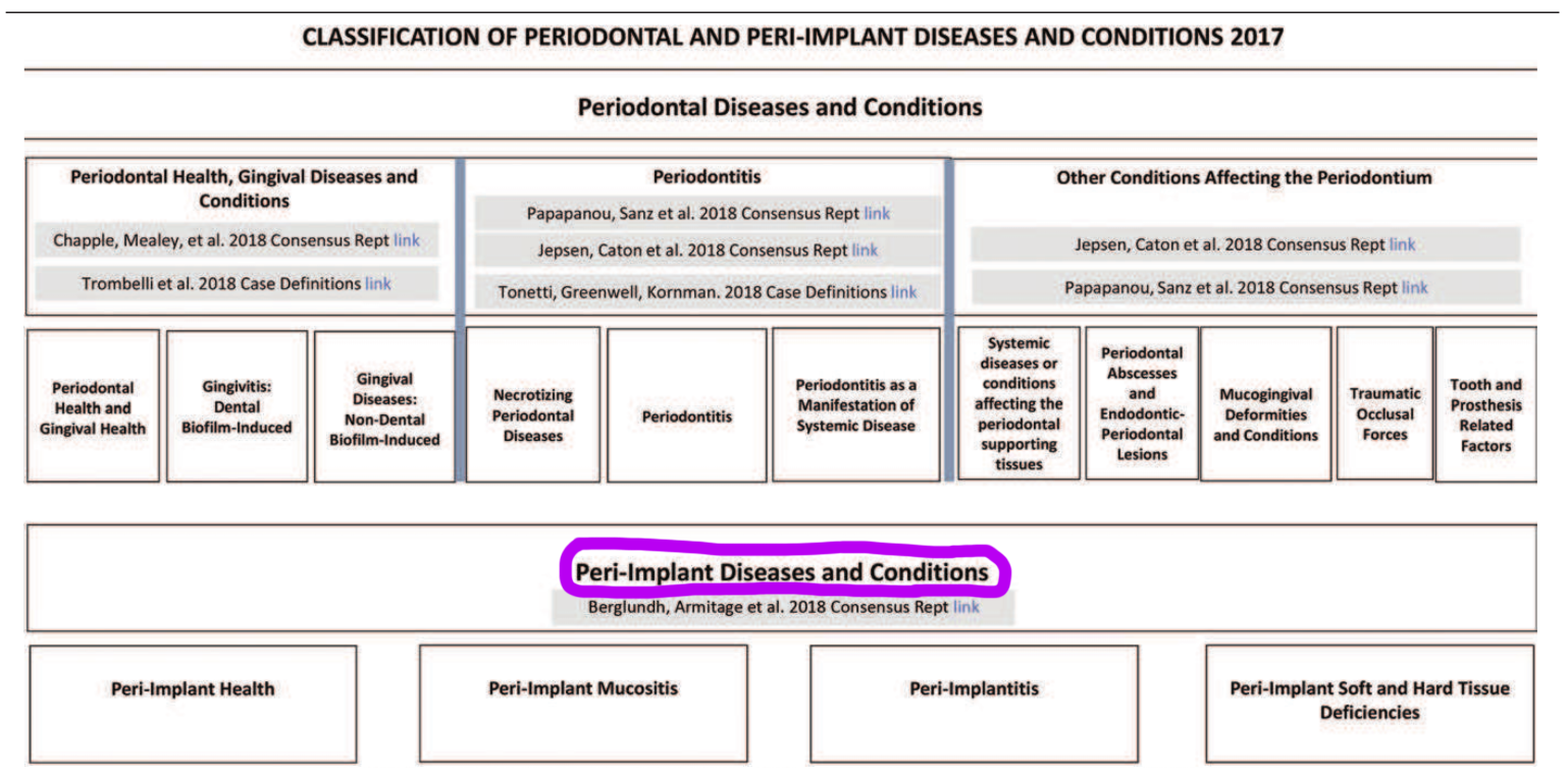
- How should you assess implants and what are the clinical characteristics of a healthy peri‐implant site?
- Visually inspect, probe and palpate to check for inflammation.
- Probe peri‐implant tissues to assess the presence of bleeding on probing, and to monitor probing depth changes and mucosal margin migration. It is not possible to define a range of probing depths compatible with health; of more importance are the clinical signs of inflammation.
- Baseline radiographic measurements following the completion of the implant supported prosthesis is recommended. An additional radiograph after a loading period should be taken to establish a bone level reference following physiological remodeling. If the patient presents for the first time with an implant‐supported prosthesis the clinician should try to obtain clinical records and previous radiographs in order to assess changes in bone levels.
- If healthy there will be no erythema, bleeding on probing, swelling and/or suppuration. There will also be no increase in probing depth compared to previous examinations. Furthermore, there should be absence of bone loss beyond crestal bone level changes resulting from initial bone remodeling.
- Peri‐implant tissue health can exist around implants with reduced bone support.
- What are the characteristics of peri-implant mucositis?
- The main clinical characteristic is bleeding on gentle probing. Erythema, swelling and/or suppuration may also be present.
- An increase in probing depth is often observed in the presence of peri‐implant mucositis due to swelling or decrease in probing resistance.
- Absence of bone loss beyond crestal bone level changes resulting from initial bone remodeling.
- What is the main aetiological factor for peri-implant mucositis?
- Plaque is the main aetiological factor but the host response to the bacterial challenge may vary between patients.
- Smoking, diabetes mellitus, and radiation therapy may modify the condition.
- Can peri-implant mucositis resolve?
- There is evidence from experimental human studies that peri-implant mucositis can resolve.
- Resolution of the clinical signs of inflammation may take more than 3 weeks following reinstitution of plaque/biofilm control.
- Peri‐implant mucositis is assumed to precede peri‐implantitis.
- What is peri-implantitis and what causes it?
- Diagnosis of peri‐implantitis requires: presence of bleeding and/or suppuration on gentle probing, increased probing depth compared to previous examinations and presence of bone loss beyond crestal bone level changes resulting from initial bone remodeling.
- Probing depth is correlated with bone loss and is, hence, an indicator for the severity of disease. Rate of progression of bone loss may vary between patients.
- In the absence of previous examination data diagnosis of peri‐implantitis can be based on the combination of: presence of bleeding and/or suppuration on gentle probing, probing depths of ≥6 mm and bone levels ≥3 mm apical of the most coronal portion of the intraosseous part of the implant.
- There is strong evidence that there is an increased risk of developing peri‐implantitis in patients who have a history of severe periodontitis, poor plaque control, and no regular maintenance care after implant therapy. Data identifying smoking and diabetes as potential risk indicators for peri‐implantitis are inconclusive. There is some limited evidence linking peri‐implantitis to factors such as post‐restorative presence of submucosal cement and positioning of implants that does not facilitate oral hygiene and maintenance. The role of peri‐implant keratinised mucosa, occlusal overload, titanium particles, bone compression necrosis, overheating, micromotion and biocorrosion as risk indicators for peri‐implantitis remains to be determined.
- Peri‐implantitis, in the absence of treatment, seems to progress in a non‐linear and accelerating pattern. Data suggest that the progression of peri‐implantitis appears to be faster than that observed in periodontitis.
- What are the main factors associated with hard‐ and soft‐tissue deficiencies at potential implant sites?
- The healing process following tooth loss leads to diminished dimensions of the alveolar process/ridge representing hard and soft‐tissue deficiencies.
- Larger deficiencies may occur at sites exposed to the following factors: loss of periodontal support, endodontic infections, longitudinal root fractures, thin buccal bone plates, buccal/lingual tooth position in relation to the arch, extraction with additional trauma to the tissues, injury, pneumatisation of the maxillary sinus, medications, and systemic diseases reducing the amount of naturally formed bone, agenesis of teeth, pressure from soft‐tissue supported removable prosthesis, and combinations.
- What factors are associated with recession of the peri‐implant mucosa?
- Malpositioning of implants, lack of buccal bone, thin soft tissue, lack of keratinized tissue, status of attachment of the adjacent teeth and surgical trauma.
- Is keratinised tissue important for the long‐term maintenance of peri‐implant health?
- The evidence is equivocal.
- It appears, however, that keratinized mucosa may have advantages regarding patient comfort and ease of plaque removal.
- What is the role of the peri‐implant bone in giving form to the peri‐implant soft tissues?
- The papilla height between implants and teeth is affected by the level of the periodontal tissues on the teeth adjacent to the implants.
- The height of the papilla between implants is determined by the bone crest between the implants.
- Results are equivocal whether the buccal bone plate is necessary for supporting the buccal soft tissue of the implant in the long‐term.
Periodontology
Recent post
June 29, 2023
Pink Aesthetics...
Read more
General Dental,Periodontology
June 26, 2022
10 Key Points from EuroPerio10...
Read more
Periodontology,Reena's Notes
June 11, 2021
Working as a hygienist or therapist...
Read more
General Dental,Periodontology