Home/Articles
/ Periodontology /
17 Key Points on Diagnostic Dilemmas in Periodontology with Prof Francis Hughes
July 7, 2016
- Periodontal diagnosis can be challenge. Identifying other pathologies particularly endodontic involvement is essential for successful treatment. It is also important to be alert for the unusual.
- 3 questions to ask yourself when forming a diagnosis: 1) What are the aetiological factors? 2) Are there any co-morbidities? 3) Is it periodontal disease?
Other pathologies – Perio-endo
- Endodontic lesions can be coincidental with periodontal problems, can cause periodontal pockets or may be caused by periodontal disease. A perio-endo lesion is a communication between the pocket and the pulp, causing a deep pocket and a non-vital tooth.
- Diagnose – radiographic most reliable, then vitality testing (isn’t always reliable and in multirooted teeth a positive response possible even if loss of vitality in part of the root canal system), pain history (frequently painless) and response to treatment. It’s important to get the diagnosis correct as if you are treating this as periodontitis alone, you will not get resolution.
- Don’t necessarily need apical involvement if lateral canal present. Also furcation lesions can rapidly lead to loss of vitality or may be as a result of a primary endodontic lesion even in the absence of apical changes.
- Diagnose the primary aetiology as this has a profound effect on the prognosis. Considerations based on: clinical judgement, periodontal disease elsewhere, reasons for loss of vitality, history/previous root treatments. Primary endo lesions often respond to RCT well, especially if of recent origin. Primary perio/true combined have a very poor prognosis and may be a good indicator for extraction.
- Management – always obturate the root canal system first or the pocket will be reinfected, then debride the affected root surface if appropriate. Debridement can be completed as soon as the root canal treated has been completed.
- Dilemma in deciding on whether or not to save the tooth. The tooth may be of strategic importance but the prognosis may be poor and the treatment extensive and expensive. Be realistic.
- Multirooted teeth – endodontic treatment and root resection of affected tooth is often extremely do-able and effective especially for upper molars (Fugazzotto 2001).
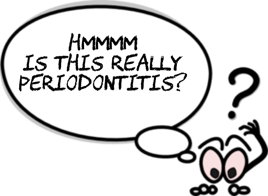
Is it periodontal disease or another condition?
- The prevalence of periodontitis is up to 45%. Other diseases of the periodontium are rare (the most common is Lichen Planus, which has a reported prevalence of 1%). So always think of the most common first – if you hear hooves, think horses, not zebras!
- The vast majority of the lesions are the results of periodontitis but these may co-exist with other conditions of the periodontal tissues or other conditions may mimic the clinical presentation of periodontitis and cause gingival redness, swelling and bone loss.
- Gingival redness – lichen planus, herpes infection, pemphigoid/pemphigus, other vesiculo-bullous disorders. Gingival swelling – leukaemia, other malignancy, Crohn’s disease, sarcoidosis, Wegener’s granulomatosis. Bone loss – Squamous cell carcinoma, other malignancies, giant cell granuloma, Langerhans’ cell histiocytosis, neutropenic conditions, hyperparathyroidism.
- Lichen planus – relatively common, soreness, redness, erosive ulcers, white lacy striae and desquamative gingivitis. Diagnosis by clinical presentation/biopsy.
- Desquamative gingivitis is a clinical description of a fiery red band on the attached gingiva, associated with lichen plus, pemphigus or other vesiculo-bullous diseases and it may coexist with gingivitis. Although it is not a cure, good periodontal care is helpful for those suffering from lichen planus (Stone et al 2015).
- Radiolucencies on your radiograph – if your periapical does not show the full extent of a lesion then this warrants further imaging. Langerhans’ cell histiocytosis can be asymptomatic and present with multiple radiolucencies in the jaw. You may miss this if the full extent of a lesion is not explored.
- Factors that should increase your suspicion – no pain, unusual appearance, change in general health, intra-oral lesion, rapidly changing lesions, not localised to the gingival margin, vital tooth within a destructive lesion, isolated single lesions in an otherwise periodontally healthy patient and unresponsive to periodontal treatment.
- Key thing is to be suspicious where appropriate and refer in good time. When referring, be specific about your concerns, offer a possible diagnosis (don’t be embarrassed to get it wrong) and ring the hospital if necessary.
These notes have been adapted from a webinar organised by the BSP. To find out about similar events and to access other resources, please visit – www.bsperio.org.uk.
Periodontology
Recent post